How to Revise Oncological Imaging and Staging Systems for the FRCR Part 2A
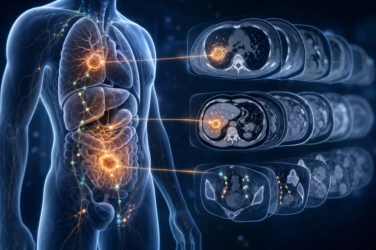
Why oncology feels so heavy
Oncological imaging has a reputation for being dry. There are staging systems to memorise, modality choices to justify, and a never-ending supply of letters and numbers. T3, N1, Barcelona B, PI-RADS 4. It can feel like you are trying to swallow a phone book.
Here is the good news. Oncology is one of the most predictable parts of the FRCR Part 2A. Examiners love it because the answers are rooted in guidelines and clear patterns. If you revise it in the right order, you stop drowning in detail and start spotting marks.
Let me walk you through how I would approach it.
Start with the principles, not the lists
Before you touch a single staging table, get the big ideas straight. These apply across almost every cancer:
- Local extent (T): what is the tumour doing where it started? Size, depth of invasion, crossing of anatomical boundaries.
- Nodal disease (N): which nodal stations drain this organ, and what makes a node look malignant?
- Metastatic spread (M): where does this cancer like to go, and what is the best test to find it?
If you understand lymphatic drainage and typical metastatic patterns, half the staging falls into place without rote learning. Prostate loves bone. Colorectal loves liver. Lung spreads to adrenals and brain. These are gifts in a single best answer question.
Learn the right modality for the right job
A huge chunk of oncology questions are really modality questions in disguise. The stem describes a cancer and asks what you would do next. You need to know the workhorse investigation for each tumour.
Quick examples worth owning cold:
- Rectal cancer: MRI for local staging, CT for distant disease.
- Prostate: multiparametric MRI, with the PI-RADS framework.
- Lung: CT first, then PET-CT for mediastinal nodes and distant disease.
- Lymphoma: PET-CT, with the Deauville score for response.
- Hepatocellular carcinoma: multiphase CT or MRI, with LI-RADS and the Barcelona staging in the background.
Make a one-line answer for every common cancer: best local test, best staging test, best follow-up test. That single habit answers a surprising number of questions.
Tackle the staging systems in small bites
Do not try to memorise a full TNM table in one sitting. It will not stick and you will hate your life. Instead, break each system into the clinically important cut-offs, because those are what change management and what examiners test.
For each major cancer, ask yourself:
- What size or depth moves it up a T stage?
- What is the first anatomical structure whose involvement upstages it?
- What node count or location matters?
- What single finding makes it unresectable?
That last question is pure gold. Encasement of the superior mesenteric artery in pancreatic cancer, for instance, is the kind of detail that turns up again and again.
Do not forget response assessment
Staging gets most of the attention, but response criteria are easy marks that many trainees skip. Know the basics of:
- RECIST: how target lesions are measured and what counts as progression or response.
- Deauville: the five-point PET scale for lymphoma.
- Choi criteria: density changes in GIST on treatment, where size alone misleads you.
You do not need encyclopaedic depth. You need to recognise the name, the cancer it applies to, and the headline rule.
Build pattern recognition with pictures
Oncology staging is visual. A T2 rectal tumour versus a T3 is a line on an MRI, not a sentence in a book. Spend time looking at annotated staging images so you can actually see where the tumour crosses a layer or breaches a capsule.
Radiopaedia is excellent for this, and working through labelled cases cements the anatomy far better than text alone. When you read about mesorectal fascia involvement, go and look at it. The picture is what you will recall in the exam.
Practise questions early and often
Reading about staging gives you a comfortable but false sense of knowing it. The only way to find the gaps is to answer questions under pressure. This is where active recall earns its keep.
This is also where SmashRad fits naturally into your revision. With over 12,000 exam-style single best answer questions, you get broad coverage of oncological imaging across every module, plus full explanations and Radiopaedia links so you can jump straight to the relevant images when something does not click. The separate Learning mode, with bite-size recall questions, is perfect for drilling those staging cut-offs and modality choices until they are automatic.
The per-module performance tracking is particularly handy for oncology, because it shows you whether your weak spot is really staging or actually the underlying anatomy. The revision recommendations then point you where to spend your limited time. Timed mock exams let you rehearse the real pressure before it counts.
A simple weekly plan
If you want a structure, try this for each organ system:
- Day one: read the principles and draw your own TNM summary from memory.
- Day two: learn the modality algorithm and response criteria.
- Day three: review annotated images for the key staging transitions.
- Day four: hammer practice questions and log every mistake.
- Revisit: come back in a week to test retention.
Spaced repetition beats cramming every single time, especially for the number-heavy content oncology throws at you.
The takeaway
Oncological imaging rewards organised revision more than raw memory. Nail the principles, own your modality algorithms, learn the staging cut-offs that change management, and keep testing yourself with pictures and questions. Do that and oncology shifts from your most feared topic to a dependable source of marks.
Ready to put it into practice? Create a free SmashRad account and try 40 sample questions, no card needed, then see how quickly your oncology confidence grows.
Put it into practice
SmashRad has 12,000+ exam-style and learning questions with full explanations and Radiopaedia links. Start free with 40 questions, no card needed.
Start free