How to Revise Breast Imaging and Mammography for the FRCR Part 2A
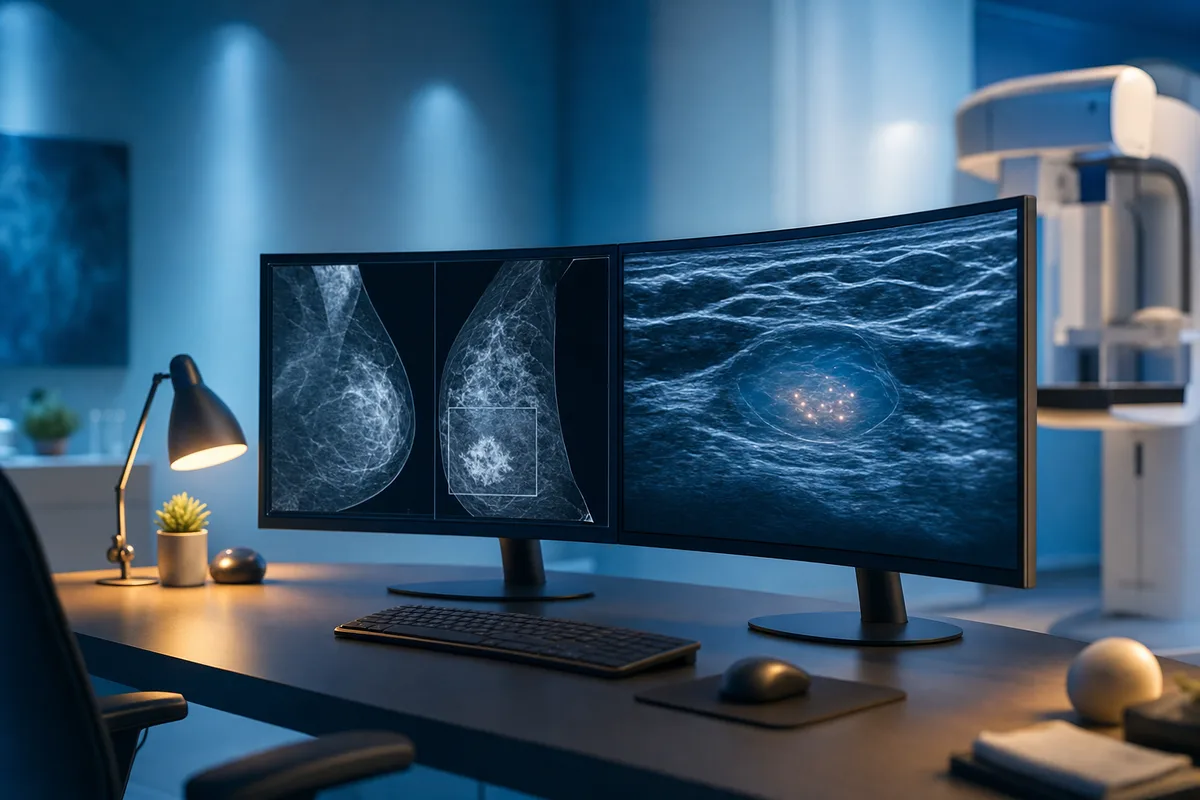
Breast imaging has a reputation among trainees. Some love it, some quietly dread it, and quite a few leave it until the very end of their revision. That is a mistake. It is a discrete, well-defined module with a predictable set of favourite topics, which means a bit of focused effort pays off nicely in the exam.
Let me walk you through how to approach it.
Why breast imaging is worth your time
The module is smaller than something sprawling like chest or musculoskeletal, but it still contributes a solid chunk of questions. Better still, the examiners tend to ask about the same core themes again and again. If you know your BI-RADS, your screening pathways and a handful of classic appearances, you can pick up marks reliably.
The catch? Mammography looks unfamiliar to many trainees, especially if your rotation has not started yet. Grainy grey images, subtle asymmetries, tiny clusters of calcification. It takes a little practice before your eye settles. So start early enough that the images stop feeling alien.
The high-yield topics
Focus your energy here first. These come up over and over.
BI-RADS and reporting
Know the assessment categories cold, from 0 through to 6, and what each one means for management. Understand breast density categories (A to D) and why density matters for sensitivity. This is pure recall, so it should be free marks once you have drilled it.
The screening programme
Understand who is invited, the age range, screening intervals and the recall process. Know the difference between screening and symptomatic pathways, and where MRI fits in for high-risk women (strong family history, known gene mutations). Questions on the two-view versus one-view approach and the role of the multidisciplinary team turn up regularly.
Calcifications
This is a classic and rewards careful study. Learn to separate benign morphology (vascular, popcorn, rim or eggshell, large rod-like, coarse) from suspicious patterns (fine pleomorphic, fine linear branching, amorphous). Distribution matters too. Grouped, linear and segmental distributions raise your suspicion. Get comfortable describing calcifications in the language the examiners expect.
Masses
Shape, margin and density are the trio to describe. Spiculated margins and irregular shapes point towards malignancy, while circumscribed oval masses lean benign. Tie this to differentials: fibroadenoma, cyst, invasive ductal carcinoma, and the trickier ones like mucinous or medullary carcinoma.
Ultrasound and MRI features
Know benign versus malignant ultrasound features (orientation, margins, posterior features, echogenicity). For MRI, understand enhancement kinetics, the type I, II and III curves, and non-mass enhancement. High-risk screening MRI is a favourite.
The must-not-miss conditions
Inflammatory breast cancer, Paget disease, radial scar, phyllodes tumour, fat necrosis and post-surgical changes all appear. Male breast disease, particularly gynaecomastia versus male breast cancer, is another reliable question.
Common traps to watch for
- Fat necrosis mimicking malignancy. Oil cysts and dystrophic calcification can look alarming. History of trauma or surgery is your clue.
- Radial scar. Spiculated on imaging but benign, though it needs sampling because of associated malignancy. Examiners love the mismatch between appearance and pathology.
- Skin thickening. Do not jump to inflammatory cancer without considering mastitis, lymphoedema, treatment change and cardiac failure.
- Overcalling benign calcification. Read the morphology carefully before you panic.
- Forgetting the second answer. Single best answer questions often list two plausible options. Pick the single most likely, not the one that is merely possible.
A revision plan that actually works
Do not try to read a whole breast textbook cover to cover. It is slow and you will forget most of it. Instead, layer your learning.
- Build the scaffolding first. Spend a short block getting the facts straight: BI-RADS, screening rules, calcification descriptors. A concise reference or a good chapter is fine here.
- Train your eye on images. Look at plenty of real mammograms and ultrasound clips. Repetition is what makes subtle findings pop out later.
- Switch to active recall as soon as possible. Reading feels productive, but answering questions is what sticks. Test yourself, get things wrong, and learn from the explanation. That loop is where real progress happens.
This is exactly where a large question bank earns its keep. On SmashRad you get over 12,000 exam-style single best answer questions with full explanations and Radiopaedia links, so when you meet a spiculated mass or an odd cluster of calcification you can read around it straight away. The separate Learning mode of bite-size recall questions is ideal for locking in the factual scaffolding like BI-RADS categories and screening criteria. The per-module tracking will also show you quickly whether breast is a strength or a gap.
Tie it together with mocks
Once you feel steady, sit timed practice under exam conditions. Breast questions sit alongside every other module in the real thing, so you need to switch systems fast without losing your rhythm. Timed mocks build that stamina and expose any lingering weak spots while there is still time to fix them.
Review every mock properly. Do not just note the score. Go through the ones you got wrong and, just as importantly, the ones you got right by luck. That honest review is worth more than another hour of passive reading.
The short version
Breast imaging is one of the more contained and predictable modules. Learn BI-RADS and screening thoroughly, get your calcification and mass descriptors precise, watch for the classic mimics, and spend most of your time doing questions rather than reading. Do that and it becomes a source of easy marks rather than a source of worry.
Ready to test yourself? Start with the free 40 sample questions on SmashRad, no card needed, and see how your breast imaging is shaping up.
Put it into practice
SmashRad has 12,000+ exam-style and learning questions with full explanations and Radiopaedia links. Start free with 40 questions, no card needed.
Start free