How to Revise Chest and Cardiac Imaging for the FRCR Part 2A
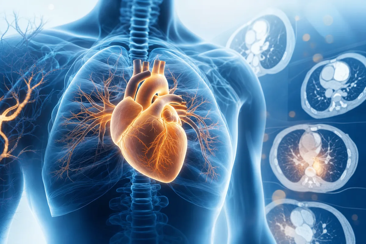
Chest imaging is the bread and butter of radiology, so it turns up everywhere in the FRCR Part 2A. Cardiac imaging is smaller but sneaky, and trainees often leave it too late. Get both right and you give yourself a solid platform for the whole exam. Here is how to revise them without drowning.
Start with the plain film, then earn your CT
It is tempting to jump straight to fancy HRCT patterns, but the chest radiograph is still where a lot of marks live. The exam loves questions that hinge on a subtle finding you either spotted or missed.
Spend real time on:
- The classic silhouette sign and lobar collapse patterns. Know which structures each lobe abuts.
- Mediastinal contours and what displaces them. Anterior, middle and posterior mediastinal masses come up again and again.
- Lines and tubes. Where should the NG tube, ET tube, central line and chest drain sit, and what does malposition look like?
- Pneumothorax, including the deep sulcus sign on a supine film.
Once the radiograph is solid, move to CT. Build a mental checklist so you review every case the same way each time. Airways, lung parenchyma, pleura, mediastinum, hila, vessels, bones and soft tissues. Consistency beats cleverness under time pressure.
Nail the interstitial lung disease patterns
ILD frightens a lot of candidates, but the exam rewards pattern recognition rather than encyclopaedic detail. Focus on distribution and the signature features.
- UIP: subpleural, basal, honeycombing, traction bronchiectasis. Think idiopathic pulmonary fibrosis.
- NSIP: subpleural sparing, ground glass, less honeycombing.
- Hypersensitivity pneumonitis: mid to upper zone, mosaic attenuation, air trapping, the classic three-density sign.
- Sarcoidosis: perilymphatic nodules, upper zone predominance, symmetrical hilar and mediastinal nodes.
Learn these four cold and you will handle most ILD questions. Then add the emergencies and infections around them.
Don't skip the acute chest
The exam mixes chronic pattern questions with acute presentations, so keep these fresh:
- Pulmonary embolism on CTPA, including signs of right heart strain.
- Aortic dissection and the Stanford classification.
- Empyema versus lung abscess (the split pleura sign is your friend).
- ARDS versus cardiogenic oedema.
A quick tip: whenever you learn a chronic condition, pair it with the acute complication that might land you in the answer options. It makes recall stickier.
Cardiac imaging: small module, big returns
Cardiac is where many trainees lose easy marks simply because they never covered it properly. You do not need to be a cardiac MRI specialist, but you do need the fundamentals.
Prioritise:
- Cardiac anatomy on CT: coronary artery segments, chamber orientation and normal calibre of the great vessels.
- Coronary artery disease: what calcium scoring means and how CT coronary angiography is interpreted.
- Congenital heart disease: the common shunts, tetralogy of Fallot, transposition and coarctation. Learn the chest radiograph appearances, like rib notching and the figure of three sign in coarctation.
- Pericardial disease: effusion, tamponade physiology and constrictive versus restrictive patterns.
- Cardiac MRI basics: late gadolinium enhancement patterns. Subendocardial equals ischaemic, mid-wall or epicardial suggests non-ischaemic causes.
That late gadolinium point alone answers a surprising number of questions, so make it second nature.
Build a high-yield fact sheet
As you revise, keep a running one-page sheet of the facts that keep catching you out. Things like the measurement thresholds for an enlarged pulmonary trunk, the CT criteria for significant lymphadenopathy, or the classic associations (lymphangioleiomyomatosis and thin-walled cysts, for example). Review this sheet in short bursts. Little and often beats one heroic session the night before.
Make questions the core of your revision
Reading is comfortable, but it lulls you into feeling prepared. The exam is single best answer, so the only way to know you are ready is to answer questions under the same conditions. Question practice does three things at once. It tests recall, it teaches you the exam's style of distractors, and it shows you exactly where your gaps are.
This is where SmashRad fits neatly into a chest and cardiac revision plan. With over 12,000 exam-style single best answer questions you will never run dry on practice, and the per-module performance tracking tells you whether your cardiac scores are quietly dragging you down. The full explanations and Radiopaedia links mean each question doubles as a mini learning session, so a wrong answer becomes a useful one.
If you prefer to build foundations first, the separate Learning mode uses bite-size recall questions to drill the facts before you attempt full cases. When you feel ready, the timed mock exams let you rehearse pacing, which matters more than most trainees expect. A free account gives you 40 sample questions with no card needed, so you can try it before committing.
A simple weekly rhythm
Tie it together like this:
- Pick a subtopic (say, ILD patterns).
- Read a focused summary or use Learning mode to lay down the facts.
- Do a block of single best answer questions on that topic.
- Log your weak areas on your fact sheet.
- Revisit those weak areas with more questions a few days later.
The spacing is the secret. You want to meet the same tricky fact several times across a few weeks, not cram it once.
Final thoughts
Chest imaging rewards structure and repetition, and cardiac imaging rewards not neglecting it. Learn your patterns, keep a tight fact sheet, and let questions do the heavy lifting of finding your gaps. Do that steadily and both modules will feel less like a threat and more like territory you own.
Ready to test yourself? Head to smashrad.com and try the free 40 questions to see where your chest and cardiac knowledge really stands.
Put it into practice
SmashRad has 12,000+ exam-style and learning questions with full explanations and Radiopaedia links. Start free with 40 questions, no card needed.
Start free